HUMB1001 · Integrated Systems Anatomy and Physiology
Module 2:
How is the Body Supported?

Bone, the skeleton and joints
→Next / continueSpacePause or resume narrationTShow transcriptNMy notesFFull screen
Sophia, the narrator of this lecture

In this lecture

  1. The skeletal system
  2. Bone as a tissue
  3. Building and growing bone
  4. The skeleton
  5. Joints
  6. Movement
Acknowledgement of Country (Boodja). I would like to acknowledge the Whadjuk Noongar people whose land we are standing on and recognise the strength, resilience and capacity of the Noongar people where Curtin University is situated. I pay my respect to their vibrant and endless culture and the leadership of the Elders past, present and future. This country (boodja) where Curtin University is situated has belonged to the Whadjuk Noongar people for thousands of years and is a place of learning for all people now and Curtin University is very proud to continue on this very long tradition.
WARNING. This material has been reproduced and communicated to you by or on behalf of Curtin University in accordance with section 113P of the Copyright Act 1968 (the Act). The material in this communication may be subject to copyright under the Act. Any further reproduction or communication of this material by you may be the subject of copyright protection under the Act. Do not remove this notice.

Press → to continue

What you'll be able to do

The skeleton's jobs
Five functions, four tissues
Bone is alive
Forming, growing, keeping calcium, repairing
Reading the skeleton
Bones by region, shape and landmark
Joints
Classified by structure and by movement
Movement
The right term for each action

A question to start

Is today's skeleton the one you'll have in ten years?

Remodelling: roughly a tenth of it, every year.

Words that build words

Word partMeansExample
osteo-, os
Bone
Osteocyte · osteon
chondro-
Cartilage
Chondrocyte · synchondrosis
peri- · endo-
Around · within
Periosteum · endosteum
-blast · -cyte · -clast
Builds · cell · breaks down
Osteoblast · osteocyte · osteoclast
syn- · arthr-
Together · joint
Synchondrosis · arthritis

Using only its word parts, what is the perichondrium?

Right. Peri means around and chondro means cartilage, so the perichondrium is the membrane wrapped around cartilage, just as the periosteum wraps around bone. You'll meet it again shortly.
Not quite. Split the word into two parts, peri and chondr. One tells you where something is, and the other tells you what it surrounds. Which option fits both?

Press → to continue

Section 1
The skeletal system

What it does, and what it's made of
Sophia, the lecture narrator

Five jobs of the skeleton

Support
A rigid framework for the soft tissues
Protection
Skull, vertebrae, rib cage
Movement
Bones as levers, turning at joints
Storage
Calcium and phosphate · fat in yellow marrow
Blood cell production
Red bone marrow

Four tissues, one system

Bone
Collagen hardened with mineral: rigid
Cartilage
Firm but flexible; smooth joint surfaces
Ligaments
Bone to bone
Tendons
Muscle to bone

A rolled ankle overstretches the tissue that holds the joint's bones together. Which tissue?

Correct. Ligaments join bone to bone, so they hold a joint together and limit how far it moves. A rolled ankle usually means a sprain: ankle ligaments that have been overstretched or torn.
Not quite. Ask what each tissue connects. One joins muscle to bone, one covers the ends of bones, and one wraps around the outside of a bone. You're looking for the one that joins bone to bone.

Press → to continue

Three kinds of cartilage

Hyaline
Smooth, glassy · joint surfaces, rib cartilages, growth plates
Fibrocartilage
Thick collagen bundles · discs, pubic symphysis, menisci
Elastic
Bends and springs back · external ear, epiglottis

How cartilage lives and grows

No blood vessels. No nerves.

Two ways to grow: on the surface, and from within.

A sports injury damages the articular cartilage on the end of the femur. How well will it heal, and why?

How confident are you?

Right. Cartilage has no blood vessels, so nutrients and repair cells can only arrive by slow diffusion through the matrix. Articular cartilage also has no perichondrium, so there's no layer of new chondroblasts on its surface. Damage here heals slowly, and often incompletely.
Not quite. Healing needs nutrients and new cells delivered to the injury. Think about what cartilage lacks that most other tissues have, and how that affects delivery. Then check whether articular cartilage has a perichondrium at all.

Press → to continue

Section 2
Bone as a tissue

Matrix, cells and structure
Sophia, the lecture narrator

Reinforced concrete

FeatureOrganic (about 35%)Inorganic (about 65%)
What it is
Collagen fibres, proteoglycans
Calcium phosphate crystals (hydroxyapatite)
What it gives
Flexibility: resists stretching and twisting
Hardness: resists compression
Remove it and…
Hard but brittle: shatters
Bends like rubber
A chicken bone soaked in vinegar can be bent, even tied in a knot. What has the vinegar removed?

How confident are you?

Right. Vinegar is an acid. It dissolves the calcium phosphate crystals but leaves the collagen behind, so the bone keeps its shape but loses its hardness. Burn a bone instead and the collagen goes: what's left is hard, but brittle enough to crumble.
Not quite. Think about which part of the matrix is a mineral and which is a protein, and which one an acid would dissolve. Then ask which part, left behind on its own, would let a bone bend.

Press → to continue

Three bone cells

Osteoblasts
Build: lay down new matrix
Osteocytes
Maintain: trapped in lacunae
Osteoclasts
Break down: dissolve matrix

Compact and spongy

Compact bone
Dense outer shell; built from osteons
Spongy bone
Trabeculae along stress lines; marrow between
Woven, then lamellar
Random collagen first, then layered sheets

Inside compact bone: the osteon

1Osteon2Central canal3Concentric lamellae4Osteocyte in lacuna5Canaliculi6Perforating canal7Circumferential lamellae + Interstitial lamellae

How bone gets its blood supply

Nutrient foramen + Nutrient arteryMedullary cavity (branches)Periosteal arteriesEpiphyseal arteriesPerforating canal + Central canalOsteocyte in lacuna + Canaliculi

Drag each structure onto the job it does.

Drag each item, or click it and then click where it goes

Lacuna
Canaliculi
Nutrient foramen
Central canal
Concentric lamellae
Perforating canal
Carries vessels lengthwise down an osteon
Carries vessels in from the surface, at right angles
Hole where the main artery enters the shaft
Small space that holds one osteocyte
Tiny channels linking osteocytes
Rings of matrix around a central canal
All six in place. The canals and the foramen deliver blood, the lacunae and canaliculi house and connect the osteocytes, and the lamellae are the matrix itself.
Not quite yet. The ones outlined in red are in the wrong place. Hole, channel, space or layer? Then ask what it holds or carries.

Press → to continue

Section 3
Building and growing bone

From embryo to adult, and for the rest of your life
Sophia, the lecture narrator

Where bone comes from: the germ layers

Ectoderm
Outer layer: epidermis, nervous system, neural crest
Mesoderm
Middle layer → mesenchyme → bone, cartilage, muscle, blood
Endoderm
Inner layer: lining of the gut and airways

Two ways to make a bone

FeatureIntramembranousEndochondral
Starts from
A sheet of mesenchyme (a membrane)
A model made of hyaline cartilage
Which bones
Flat skull bones, part of mandible, clavicle shaft
Most others: limbs, vertebrae, pelvis, skull base
How
Mesenchyme cells become osteoblasts directly
Cartilage is replaced by bone
First bone made
Woven, later remodelled to lamellar

Endochondral ossification

1Cartilage model
2Bone collar; cartilage calcifies
3Primary ossification centre
4Medullary cavity forms
5Secondary ossification centres
6Plate and joint surfaces remain

The epiphyseal plate: five zones

Resting cartilage
Anchors the plate to the epiphysis
Proliferation
Chondrocytes divide, stacking in columns
Hypertrophy
Chondrocytes swell
Calcification
Matrix calcifies; chondrocytes die
Ossification
Osteoblasts lay bone on calcified cartilage
Growth stops
Plate replaced by bone → epiphyseal line

On a child's knee X-ray, a dark band crosses each end of the femur. Why is it dark?

Right. X-rays are blocked by mineral, so bone shows up white and soft tissue, including cartilage, looks dark. The dark band is the epiphyseal plate, so this child is still growing. In an adult, you'd see only a thin white epiphyseal line.
Not quite. On an X-ray, mineralised tissue shows up white and everything else looks dark. Which tissue is still found across the ends of a child's long bones, and is it mineralised?

Press → to continue

What shapes growth

FactorRole in boneWhen it goes wrong
Calcium and protein
Mineral and collagen; calcium ~1,000 mg/day (1,300: teens, women 50+, men 70+)
Smaller, weaker bones
Vitamin D
Lets the gut absorb calcium
Soft bones: rickets (children), osteomalacia (adults)
Vitamin C
Needed to make collagen
Scurvy: weak bone, slow healing
Growth and thyroid hormones
Drive growth at the plates
Too little in childhood: short stature
Sex hormones
Puberty growth spurt, then plate closure
Growth stops once the plates close

Calcium balance: PTH and calcitonin

1Blood calcium falls
2PTH released
3Bone: PTH acts on osteoblasts → osteoclasts
4Kidneys keep calcium; make active vitamin D
5Gut absorbs more calcium
6Back in range → PTH falls (if too high: calcitonin)
PTH raises blood calcium in three ways. Which of these is NOT one of them?

How confident are you?

Right. PTH does the opposite: it makes the kidneys hold on to calcium. The other three are PTH's three routes, and remember that the bone route is indirect, with PTH acting on osteoblasts, which then switch on osteoclasts.
Not quite. Go back through the three places PTH acts: bone, kidneys and gut. For each option, ask whether it would push blood calcium up or down. Only one of them would lower it.

Press → to continue

Bone is rebuilt all the time

Out with the old
Osteoclasts remove, osteoblasts rebuild
Load builds bone
Denser bone; trabeculae along stress lines
Disuse loses bone
Bed rest, a plaster cast, spaceflight

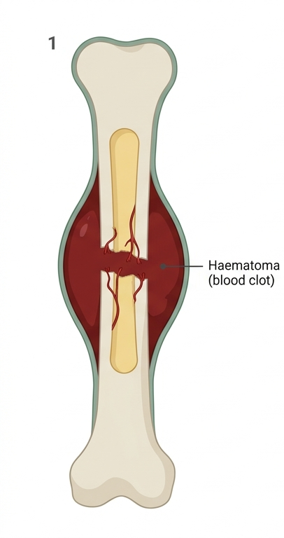
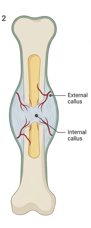
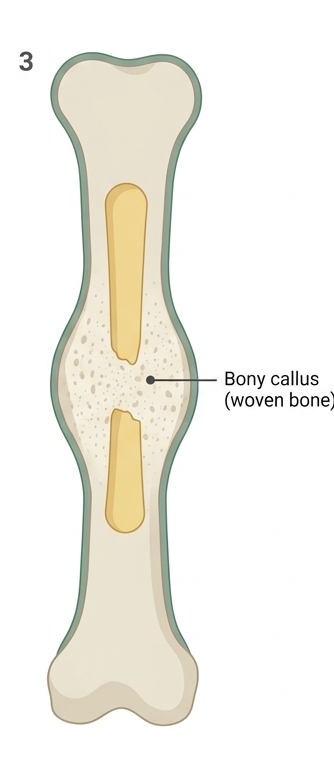
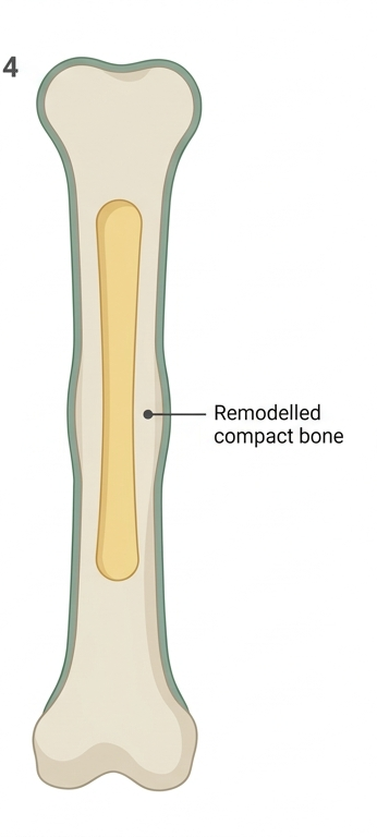
Put the stages of fracture repair in order.

Drag the steps into order, first at the top, or use the arrow buttons.

  1. ⋮⋮Lamellar bone replaces the woven bone
  2. ⋮⋮Fibrocartilage and fibrous tissue bridge the gap
  3. ⋮⋮A blood clot fills the break
  4. ⋮⋮Woven bone replaces the callus
That's the sequence: haematoma, callus, bony callus, remodelling. The callus is first made of cartilage and fibrous tissue, and woven bone comes before lamellar bone, just as it does in the embryo.
Not quite. The first thing a break does is bleed. Then think about which tissue is quickest to make, and which kind of bone always comes first: woven or lamellar?

Press → to continue

Section 4
The skeleton

Naming bones, and reading their surfaces
Sophia, the lecture narrator

Axial and appendicular

Axial: skull
Cranium and face, plus ear ossicles and hyoid
Axial: trunk
Vertebral column, ribs and sternum
Appendicular: upper
Pectoral girdle and upper limbs
Appendicular: lower
Pelvic girdle and lower limbs

Tour the skeleton

1 2 3 4 5 6 7

Click a region to see the bones you need to know there.


Bones to know

–

Tip

–

0 of 7 explored

Four shapes of bone

Long
Longer than wide: limbs, fingers, toes
Short
About as wide as long: wrist and ankle
Flat
Thin plates: skull roof, ribs, sternum, scapula
Irregular
Complex shapes: vertebrae, some facial bones

Anatomy of a long bone

1Diaphysis2Proximal epiphysis + Distal epiphysis3Articular cartilage4Epiphyseal line5Medullary cavity (yellow marrow)6Red marrow (in spongy bone)7Periosteum8Endosteum

Reading the landmarks

FamilyJobTerms
Projections
Anchor muscles, tendons and ligaments
Process, tubercle, tuberosity, trochanter, crest, spine, epicondyle, line
Articular surfaces
Form a joint with another bone
Head, condyle, facet
Depressions and openings
House structures or let them pass
Fossa, groove, notch, foramen, canal, meatus, fissure

Worked example: the femur

1Head + Fovea capitis2Neck3Greater trochanter4Lesser trochanter5Intertrochanteric line + Intertrochanteric crest6Gluteal tuberosity7Linea aspera + Nutrient foramen8Medial condyle + Lateral condyle + Patellar surface9Medial epicondyle + Lateral epicondyle + Adductor tubercle10Intercondylar fossa
1 2 3 4 5 6 7 8 9 10

Click the feature whose name tells you it's a depression.

Click a numbered marker.

Sort each feature into its family.

Drag each item, or click it and then click where it goes

Foramen magnum
Olecranon fossa
External acoustic meatus
Spinous process of a vertebra
Head of the humerus
Tibial tuberosity
Facet on a thoracic vertebra
Condyle of the mandible
Iliac crest
Projection
Articular surface
Depression or opening
All nine sorted. The feature word carries the clue every time: crest, tuberosity and process stick out; head, condyle and facet form joints; foramen, meatus and fossa are holes, tunnels or hollows. (The external acoustic meatus is also called the external auditory meatus.)
Not quite yet. The ones outlined in red are in the wrong place. Ignore the bone. Look at the feature word: which family does it belong to?

Press → to continue

Section 5
Joints

Where bones meet, and how much they move
Sophia, the lecture narrator

Two ways to classify a joint

Structural classWhat joins the bonesHow much it usually moves
Fibrous
Dense fibrous connective tissue; no cavity
Immovable or slightly movablesynarthrosis or amphiarthrosis
Cartilaginous
Cartilage; no cavity
Immovable or slightly movablesynarthrosis or amphiarthrosis
Synovial
A fluid-filled cavity inside a capsule
Freely movablediarthrosis

Fibrous joints

Sutures
Interlocking skull bonesImmovable – synarthrosis
Syndesmoses
Bones joined by ligament or membraneSlightly movable – amphiarthrosis
Gomphoses
A tooth in its socketImmovable – synarthrosis

Cartilaginous joints

Synchondroses
Hyaline cartilageMostly immovable – synarthrosis
Symphyses
A pad of fibrocartilageSlightly movable – amphiarthrosis

What type of joint is a child's epiphyseal plate, and what does it become in an adult?

Right. The plate joins the epiphysis to the shaft with hyaline cartilage, so it's a synchondrosis. When growth stops, the cartilage is replaced by bone and the two parts fuse: a synostosis, which is the epiphyseal line you saw on the adult femur.
Not quite. First ask what kind of cartilage the plate is made of, and which joint type uses it. Then think back to what happens to the plate when growth stops. What replaces the cartilage?

Press → to continue

Anatomy of a synovial joint

Freely movablediarthrosis

1Articular cartilage2Joint cavity (synovial fluid)3Synovial membrane4Fibrous capsule5Ligament6Bursa7Tendon

Six shapes of synovial joint

Plane
Flat surfaces slide · between carpals and tarsals
Hinge
One plane, like a door · elbow, knee
Pivot
Rotation around one axis · atlas and axis, radius
Condyloid
Oval in an oval · wrist, knuckles
Saddle
Two saddles fit together · base of the thumb
Ball-and-socket
Every direction, including rotation · shoulder, hip

Shoulder vs hip

FeatureShoulderHip
The socket
Shallow glenoid cavity; large head
Deep acetabulum grips the head
Capsule and ligaments
Loose capsule; relatively weak ligaments
Thick capsule; very strong ligaments
Muscles
Rotator cuff tendons hold the head in place
Powerful muscles all around add support
The result
Most mobile joint; dislocates most often
Very stable; rarely dislocates

Sort each joint into its structural class.

Drag each item, or click it and then click where it goes

Knee
Shoulder
Tooth in its socket
Interosseous membrane between radius and ulna
Child's epiphyseal plate
Elbow
Intervertebral disc
Coronal suture
Pubic symphysis
Fibrous
Cartilaginous
Synovial
All nine sorted. Sutures, gomphoses and syndesmoses are fibrous; synchondroses and symphyses are cartilaginous; and every freely movable joint with a fluid-filled cavity is synovial.
Not quite yet. The ones outlined in red are in the wrong place. Ask what joins the bones: fibrous tissue, cartilage, or a fluid-filled cavity?

Press → to continue

Section 6
Movement

Naming what joints do
Sophia, the lecture narrator

The reference point

Every movement starts from the anatomical position.

Most come in opposite pairs. Two reference planes: coronal and median.

Angular movements

1Flexion and extension
2Hyperextension
3The knee is the exception
4Plantar flexion and dorsiflexion
5Abduction and adduction
6Fingers and toes

Circular movements

1Rotation
2Pronation and supination
3Circumduction

Special movements

1Elevation and depression
2Protraction and retraction
3Excursion
4Opposition and reposition
5Inversion and eversion

Movements at selected joints

1 2 3 4 5 6 7

Click a joint to see its type and the movements it allows.


Joint type

–

Movements

–

0 of 7 explored

Drag each action onto the movement it involves.

Drag each item, or click it and then click where it goes

Bending at the hip as you sit down
Turning your palm up to receive change
Pushing your jaw forward
Touching your thumb to your little finger
Shrugging your shoulders
Standing on tiptoe
Elevation
Plantar flexion
Supination
Opposition
Protraction
Flexion (at the hip)
All six matched. Shrugging lifts the scapulae, tiptoe points the foot towards the sole, supination turns the palm up, opposition brings the thumb across the palm, protraction glides the jaw forward, and sitting bends the hip forward of the coronal plane.
Not quite yet. The ones outlined in red are in the wrong place. Do the action yourself, then ask which way the part moves and which pair that movement belongs to.

Press → to continue

Flex your shoulder to 90°, flex your elbow to 90°, then fully pronate your forearm. Which way does your palm face?

How confident are you?

Right. Flexing the shoulder points your arm forward, and flexing the elbow points your forearm up, so your palm faces back towards your face. Full pronation then turns the palm half a turn, so it faces forwards, away from you.
Not quite. Do it step by step, starting from the anatomical position with your palm facing forward. Notice which way your palm faces after each step, then remember that pronation turns the palm over, half a turn.

Press → to continue

Five big ideas

Bone is alive
Cells in a mineral-and-collagen matrix, with a blood supply
Built on a model
Cartilage first; growth at the plate until it closes
A calcium bank
PTH draws on bone; remodelling never stops
The name gives the job
Projections, articular surfaces, depressions and openings
Structure decides movement
Joint type and shape set the range

Reflect

1. A young adult spent their whole childhood in a refugee camp. Would you expect them to reach their potential height? Explain why or why not.

Saved on this device

2. Choose one idea from this lecture that will matter in your future profession, and explain how.

Saved on this device

Your answers are saved only in this browser. Nobody else can see them. They appear on your notes page next.

Your notes

HUMB1001 · Module 2 · How is the Body Supported?

Five big ideas

  1. Bone is alive — Cells in a mineral-and-collagen matrix, with a blood supply
  2. Built on a model — Cartilage first; growth at the plate until it closes
  3. A calcium bank — PTH draws on bone; remodelling never stops
  4. The name gives the job — Projections, articular surfaces, depressions and openings
  5. Structure decides movement — Joint type and shape set the range

My reflections

A young adult spent their whole childhood in a refugee camp. Would you expect them to reach their potential height? Explain why or why not.

Choose one idea from this lecture that will matter in your future profession, and explain how.

Before your lab this week

  • Seeley chapters 6–8
  • Pre-lab “The Skeleton”; print your lab notes
  • Lab coat, safety glasses, closed-in shoes; long hair tied back
  • Anatomy Compliance Quiz at 100% → authorisation sticker
  • After the lab: Module 2 revision quizzes and FeedForward questions